CBCT Scans
Dental cone beam computed tomography (CT) is a special type of x-ray machine used in situations where regular dental or facial x-rays are not sufficient. This type of CT scanner uses a special type of technology to generate three dimensional (3-D) images of dental structures, soft tissues, nerve paths and bone in the craniofacial region in a single scan. Images obtained with cone beam CT allow for more precise treatment planning.
You should wear comfortable, loose-fitting clothing to your exam. You may be given a gown to wear during the procedure.
Metal objects, including jewelry, eyeglasses, dentures and hairpins, may affect the CT images and should be left at home or removed prior to your exam. You may also be asked to remove hearing aids and removable dental work. You may be asked to remove any piercings, if possible.
Cone beam CT is not the same as conventional CT. However, dental cone beam CT can be used to produce images that are similar to those produced by conventional CT imaging.
With cone beam CT, an x-ray beam in the shape of a cone is moved around the patient to produce a large number of images, also called views. CT scans and cone beam CT both produce high-quality images. Dental cone beam CT was developed as a means of producing similar types of images but with a much smaller and less expensive machine that could be placed in an outpatient office.
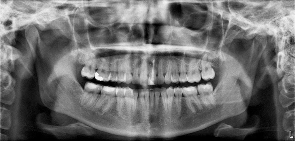
Cone beam CT provides detailed images of the bone and is performed to evaluate diseases of the jaw, dentition, bony structures of the face, nasal cavity and sinuses. It has the advantage of lower radiation exposure compared to conventional CT.
Dental cone beam CT is commonly used for treatment planning of orthodontic issues. It is also useful for more complex cases that involve:
- surgical planning for impacted teeth.
- diagnosing temporomandibular joint disorder (TMJ).
- accurate placement of dental implants.
- evaluation of the jaw, sinuses, nerve canals and nasal cavity.
- detecting, measuring and treating jaw tumors.
- determining bone structure and tooth orientation.
- locating the origin of pain or pathology.
- cephalometric analysis.
- reconstructive surgery.*
* from radiologyinfo.org